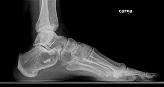
Imagen radiográfica:
Se observa lesión lítica en la región anterior del calcáneo con una calcificación en su interior:
Imagen del Tac realizado donde se observa la lesión lítica con una densidad ósea en su interior muy sugerente de lipoma intraóseo.
Clasificación de Milgran de los lipomas intraóseos
Milgram JW. Intraosseous lipomas. A clinicopathologic study of 66 cases. Clin
Orthop 231:277–302, 1988.
Grupo I lesiones puramente liticas con lipocitos maduros y quiste cubierto de fina esclerosis sin signos de necrosis,
grupo II lesiones parcialmente radiotransparentes con lipocitos mezclados parcialmente necroticos y viables,
grupo III las cuales presentarian una gran clacificación central y periférica sin trabéculas óseas en su interior y lipocitos completamente necroticos
Revisión de los quistes de calcáneo, donde se define el tamaño crítico de los mismos:
Clin Orthop Relat Res. 2004 Jul;(424):202-10.
Clinical relevance of calcaneal bone cysts: a study of 50 cysts in 47 patients.
Pogoda P, Priemel M, Linhart W, Stork A, Adam G, Windolf J, Rueger JM, Amling M.
SourceDepartment of Trauma, Hand, and Reconstructive Surgery, Hamburg University School of Medicine, Marti...
nistrasse 52, 20246 Hamburg, Germany.
Abstract
The clinical relevance and nature of calcaneal cysts is controversial. The risk of pathologic fracture is undefined and diagnostic criteria to differentiate between cysts in patients who can be treated nonoperatively and patients who require surgical intervention are not available. To address these questions, 50 calcaneal bone cysts in 47 patients were evaluated. The majority of cysts (40 of 50) were asymptomatic and were treated nonoperatively. Cysts reaching a critical size, defined as 100% intracalcaneal cross section in the coronary plane and at least 30% in the sagittal plane, are at risk for becoming symptomatic and at risk for fracture. Fracture is a significant complication and occurred in four of 47 patients, three of whom were treated by open reduction internal fixation and bone grafting. In addition, six patients with symptomatic critical size cysts without apparent fracture were treated by curettage and subsequent autogenous bone grafting or calcium-phosphate cement filling, and there were no recurrences. We report one of the largest series of cysts in the calcaneus. The results suggest that calcaneal cysts are clinically relevant because of the potential risk of fracture and that size is a significant factor in terms of the treatment of the cyst.
En resumen un quiste que alcanza el 100% del espesor en sentido medio lateral y un 30% en anteroposterior alcanza e tamaño critico susceptible de sufrir una fractura patológica y estaría indicado intervenirlo.
Lipoma intraóseo del calcáneo. A propósito de un caso clínico :
http://vitae.ucv.ve/?module=articulo&rv=101&n=4440&m=3&e=4476
En el Journal on Bone Surgery BR se revisa el beneficio de realizar el curetaje de manera artroscópica:
J Bone Joint Surg Br. 2011 Dec;93(12):1626-31.


No hay comentarios:
Publicar un comentario